Update: 21 December 2024
Toxicovigilance
Persistent environmental exposure to toxic chemicals, metals, microplastics and technologies such as electromagnetic fields harm everyone. Our environment is now a leading cause of cancer. The World Health Organization estimates that 85% of all human disease is linked to environmental toxicity.

Poisonous air is having a devastating impact on billions of children around the world, damaging their intelligence and leading to hundreds of thousands of deaths. According the WHO – 1.8 billion children – more than 90% of the world's young people are breathing toxic air, creating a public health time bomb.
Contents
- Toxicovigilance in a Toxic World
- Microplastic Ingestion
- What can you do about it?
- Toxic Heavy Metals
- Common Toxic Metals
- Biomonitoring - Testing for Heavy Metals
- Resolving Toxicity
- Resources
Toxicovigilance in a Toxic World
Every minute of everyday we are exposed to toxicity. Toxins are in the air we breathe and the food we eat. Environmental illness is the inevitable consequence of exposure to toxins from a multitude of sources including, chemicals, pharmaceuticals, coal smoke, mining operations, automobiles, landfills, fertilizers, pesticides herbicides and fungicides, even drinking water is toxic.
A new study of pregnant women and their newborns find more than 50 new – never observed in people before – environmental chemicals. For most, the health consequences are unknown. These findings are concerning, as pregnant women and newborns are most vulnerable.
- More Than 50 New Environmental Chemicals Detected in People Ashley P. Taylor
‘In today's world, everyone carries a toxic load of dozens of industrially produced chemicals in their bloodstream. Not only do these adversely affect the health of adults and children, but also, and more worryingly, they damage the development of unborn infants. The amniotic fluid of pregnant women has been found to contain a variety of chemicals, such as pesticides, plasticizers, disinfectant products, flame-retardants, surfactants and UV filters, many of which interfere with fetal physiology, especially thyroid hormone action.
Thyroid hormone is vital for brain development, particularly for the fetus during pregnancy and for toddlers. In fact, children born to women who lack this thyroid hormone (or who are unwittingly exposed to thyroid-disrupting chemicals) have lower IQs and more neurodevelopmental problems.’ Barbara Demeneix, Toxic Cocktail: How Chemical Pollution Is Poisoning Our Brains
The air we breathe is causing brain injury for millions.
The American Lung Association reports — almost half of the US population (139 million) — live in areas where "the air is too dangerous to breathe." A growing body of evidence suggests that pollution harms the brain. New research connecting neurodegenerative diseases like Alzheimer's and Parkinson's to the dirty air we breathe shows that pollution may also age the brain prematurely. According University of Texas-El Paso researchers, pollution's damage to the brain may start even sooner. Fourth and fifth grade children who are exposed to toxic air pollutants at home are more likely to have lower GPAs.
Microplastic Ingestion
Microplastics are contaminating the air we breathe, the food we eat, and the water we drink. Microplastics are defined as plastic particles under 5mm in size. Primary microplastics are plastics directly released into the environment in the form of small particulates (shower gel microbeads, tire abrasion, etc.) while secondary microplastics are microplastics originating from the degradation of larger plastic (e.g. degraded plastic bags).
The average person is unintentionally ingesting approximately 5 grams of plastic per week (the equivalent of eating a credit card).
Ingestion is just one aspect of a much wider plastics crisis. Plastic pollution is a major threat to wildlife, not only through microplastic ingestion but via entanglement and habitat destruction. Plastic pollution also has damaging economic consequences, with the UN Environment Programme (UNEP) estimating its annual economic impact on the ocean economy at US$8 billion.
- Assessing Global Plastic Ingestion ©WWF – World Wide Fund For Nature, Gland, Switzerland
What can you do about it?
We are largely kept ignorant about the magnitude and seriousness of toxicity issues. Successful lobbying efforts have resulted in self-protective policy and laws for some of the corporations responsible. They also use well-executed (dis)information campaigns to misdirect public awareness and understanding.
But we the people can have an immediate, powerful impact when we withdraw our support. Stop buying their products - our money is their life blood. We can render them irrelevant.
You can personally manage the problem with your own toxicovigilance. Avoid and reduce your exposure to toxins, support your body's stress response and detoxification function, fortify your immune system. And you can verify detoxification to ensure progress. Biomonitoring agencies worldwide use hTMA testing as the standard protocol for identifying toxicity and monitoring the detoxification. The U.S. EPA also recommends hTMA as the ‘testing method of choice’ for determining and monitoring chronic heavy metal poisoning.
Detoxification is tricky business and can be life threatening if aggressive chelation therapies are used. We recommend that you work closely with a qualified hTMA practitioner to ensure safety and effectiveness.
Back to TopToxic Heavy Metals
Toxic metals are abundant in the environment. Many of common heavy metals are directly toxic to cells. For example, the presence of mercury in the body is always a bad thing. When essential nutrient minerals are not available in adequate amounts, available toxic metals are often substituted in cellular binding sites. For instance, lead is a substitute for calcium if the calcium element is unavailable. When the levels of toxic metals are too high in the body's soft tissues, they will displace essential nutrient minerals. This kind of mineral substitution and displacement leads to cellular dysfunction, illness, disease, even death.
Heavy Metal Accumulation
Heavy metals can enter the body through inhalation, intestinal absorption and even be absorbed through the skin, depending upon their chemical form. Elemental forms of heavy metals are not well absorbed, but organometallic forms are lipophilic, and can readily pass through membranes and even cross the blood-brain barrier (our neurological defense system).

Once absorbed into the body, heavy metals have a wide distribution in various organs, glands and the central nervous system. Some metals, like lead, are bone seekers and ultimately settle into the teeth and skeletal system. Heavy metals then can effectively poison enzyme systems, increase free radical production and displace or compete with essential elements that make up metallo-enzyme complexes and compete with the absorption of nutritional minerals.
Toxin levels in body tissues increase with every generation, even when exposure levels stay the same. This is because the growing fetus is far more vulnerable than the adult and most toxins pass through the placenta in-utero. A female child born to a mother who has a copper excess will begin life with a higher copper level than her mother began life. Throughout her developing years, such a female child is likely to accumulate more and more copper in her tissues, especially during pre-adolescence and adolescence when her own estrogen production increases. She is likely to give birth to children who will absorb higher amounts of copper than she absorbed in utero. In each succeeding generation, this cycle is likely to repeat itself resulting in more and more children born with significant copper excesses (see graph). This process of transmitting more and more copper in utero from one generation to the next with increasing amounts of excess copper building up and accumulating to toxic levels in each generation is likely to produce an epidemic of psychological and physical problems associated with excess tissue copper. This is primarily due to the fact that excess copper tends to be stored in the brain and in the liver.
The increasing numbers of children and adolescents who have learning and behavior problems can be partly related to the accumulation and transmission of excess copper from one generation to the next. Such a trend has very serious implications for families, schools, and society in general. The incidence rates of Learning Disabilities and Attention Deficit Disorder are likely to continue to increase as excess copper is transmitted from one generation to the next. Aggressive and violent behavior also will tend to increase with the build up of excess tissue copper levels. Addictive cravings and addictive behavior also are likely to increase. Depression and panic attacks will likely also increase in frequency.
Back to TopCommon Toxic Metals
One of the major contributors to health problems is the constant exposure to harmful toxins prevalent in our environment. Some of the most physically and neurologically damaging of these are the metals; aluminum, lead, mercury, arsenic, and cadmium. They contribute to everything from teeth problems and bone disease, to worsening allergies and skin diseases, to serious life threatening illnesses such as cancer, arthritis, kidney failure, congestive heart disease, liver diseases and diabetes. Heavy metals damage the immune system, reducing defenses against infections as well as bacterial, viral and fungal diseases.
- Aluminum
-
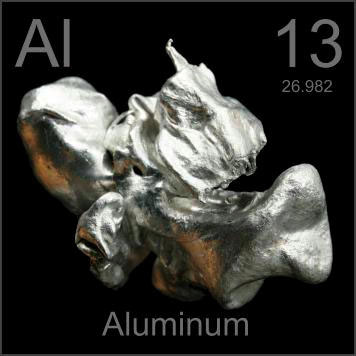
Aluminum has been described as a protoplasmic poison and a pernicious and persistent neurotoxin. While the body is able to excrete aluminum in its natural form, the element, like mercury, is toxic to all life forms when concentrated in their tissues. It has a tendency to accumulate in the brain and nerve tissues and in the bones and teeth.
Aluminum interferes with the absorption of a number of essential elements including, iron, fluoride, phosphorus and calcium. It inhibits gastric muscle contraction and can cause constipation. This disrupting effect on the essential minerals leads to endocrine gland dysfunctions as these glands all depend on balanced mineral ratios. These dysfunctions include hypo- or hyperthyroidism, hypo- or hyper-adrenal, hypoglycemia, diabetes, dry dull coats, dry or flaky skin, and digestive disorders due to lack of pancreatic digestive enzymes and lowered stomach hydrochloric acid.
Aluminum is a neurotoxin. Central nervous system symptoms found in individuals with aluminum toxicity include dementia, aggression, and violent behavior.
Aluminum's toxic effect on the skeletal system was first recognized in the late 1970s. In animal studies it has been found to induce anemia. Any impairment of kidney function will increase aluminum toxicity as the kidneys are the main route of excretion.
Aluminum can impair cellular energy transfer processes by interfering with phosphate and ATP metabolism. Since aluminum has an affinity for brain and nerve tissue, it can affect any organ in the body via the central nervous system. This can lead to a multitude of health problems and a weakening of the immune response. Aluminum toxic individuals are more susceptible to bacterial, fungal and viral diseases, and chronic dermatitis. Other symptoms of aluminum toxicity include extreme nervousness, weak muscles, seizures, loss of balance, and loss of energy.
- Lead
-
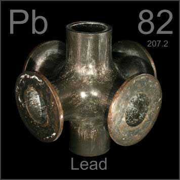
Lead can be found in all parts of our environment. Much of it comes from human activities including burning fossil fuels, mining, and manufacturing. The concentration of lead in polluted air varies inversely with altitude. Because lead is a heavy element, it settles out of the air onto the ground. We are exposed to lead from aviation fuel, paint on older buildings, lead pipes, many industrial uses, coal burning, and pesticides which can contain high amounts of lead. Lead is used in the seams of canned food cans where it leaches into the food. Lead can even be in your drinking water.
Calcium inhibits lead absorption and protects against lead accumulation in bones and teeth. Young children are naturally fast metabolizers with lower levels of tissue calcium and magnesium. This low calcium to lead ratio leads to higher lead absorption into the tissues. Common storage sites for lead are bones and teeth, where it displaces calcium. During times of stress, when the body would normally release more calcium (a calming mineral), it will release stored lead into the bloodstream. This can bring lead into any of the body's organs, leading to cancer and organ failure.
Besides calcium, lead also antagonizes and prevents the absorption of other important nutrient minerals such as iron and zinc. Lead interferes with iron metabolism and contributes to anemia. Other nutrients such as magnesium, copper, chromium, vitamin C and the B vitamins have been shown to protect against the effects of lead as well, either by decreasing its absorption and tissue deposition, or by reducing or blocking the effects of lead upon enzyme systems. hTMA will verify what your tissue lead levels are and whether there are adequate amounts of the protective minerals present to prevent lead accumulation in the tissues.
Epilepsy can result from lead toxicity. In young children, hyperactivity may be the first presenting symptom. Other emotional symptoms of a lead body burden include aggression, violent behavior, antisocial behavior, poor concentration or learning capacity. It can take years before low-level lead exposure reaches dangerous levels, so you might not recognize symptoms until a child is older. Since lead can block and prevent many enzyme functions, lead toxicity can lead to damage to the heart, kidneys, liver, gastrointestinal tract, and central nervous system. Symptoms of lead toxicity include lack of appetite, vomiting, abdominal pain, constipation followed by diarrhea, crunching of jaws, blindness, seizures or muscle spasms, behavior changes, and loss of balance and agility.
- Mercury
-
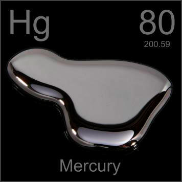
Mercury is one of the most studied toxic heavy metals and the lethal effects of both acute toxic exposure and chronic low-level exposures are well documented. Exposure to mercury occurs from breathing contaminated air, ingesting contaminated water and food, and having dental and medical treatments. Mercury can damage the brain, kidneys, and developing fetus. This chemical has been found in at least 714 of 1,467 National Priorities List sites identified by the Environmental Protection Agency. We are all told to avoid eating too much fish, and pregnant women are told to avoid it completely due to the problems that mercury causes to the growing fetus. For all of human history, fish was always one of the best and most nutritious foods we could eat.
Methylmercury is the preservative used in most vaccines, and can be found in some pharmaceutical medications. It is used in household products such as batteries, light bulbs, fabric softeners, latex gloves, paint, plastics, ink, and solvents. Mercury vapors are released from home renovations involving old paint, broken thermometers and thermostats. Even some cosmetics contain mercury. Mercury salts are sometimes used in skin lightening creams and in antiseptic creams and ointments. Like other heavy metals, it is in our air, water and on the ground. We are all continually exposed.
Mercury prevents glucose transport, thus reducing cellular energy availability. Mercury antagonizes and prevents the absorption of important nutrient minerals such as zinc, iron, selenium and sulfur. hTMA analysis will verify what your tissue mercury levels are and whether there are adequate amounts of the protective minerals present to prevent mercury accumulation in the tissues.
Mercury accumulates in the brain and central nervous system. Mercury also adversely affects your overall immune system by attaching to the immune cell structure and altering their ability to function normally. Mercury can cause kidney and cardiac diseases, respiratory problems, arthritis, and gum disease.
Symptoms of high mercury tissue levels include loss of balance, fatigue, vomiting, hair loss, diarrhea, weakness, and excessive salivation. High levels also interfere with enzyme activity, and can result in blindness and paralysis. Mercury causes convulsions, anorexia, tremors, swollen gums and behavior problems.
- Arsenic
-
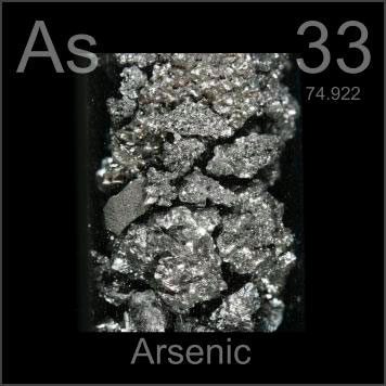
Arsenic is public enemy number one on ATSDR's Priority List of Hazardous Substances list. (Lead is #2, mercury is #3.) Arsenic is a known carcinogen and affects the skin, digestive system, liver, nervous system and respiratory system. Arsenic compounds can create reactions in the body that disrupt enzymes that are involved in respiration of cells, fat and carbohydrate breakdown and their metabolism. The accumulation of toxic levels of arsenic can result in paralysis, coma, cardiovascular collapse and death.
Today in the United States, the quantity of arsenic released by human activities exceeds amounts released from natural sources by at least threefold. Major sources of arsenic release into the environment are;
- The Mining and Chemical Industries.
- Coal fired power plants.
- Confined Animal Feeding Operations (CAFOs).
- Arsenic-treated lumber.
In modern industrial agriculture (i.e., CAFOs) arsenic is fed to pigs and poultry to improve production. In the 1940s producers began using the organic form of arsenic to promote growth, treat disease and improve meat pigmentation. By 2010, according to industry estimates, 88% of chickens raised for human consumption in the U.S. were given the arsenic-based feed/drugs. In the case of swine, arsenic is used to treat diarrhea. This meat is then used in commercial food products. Arsenic has been found in at least 1,149 of the 1,684 National Priority List sites identified by the Environmental Protection Agency. Also, arsenic can be found in many commonly used products including fungicides, pesticides, herbicides, laundry products, cigarette smoke, paints, and wood preservatives. Global industries such as mining and smelting, chemical and glass manufacturing produce arsenic as a by-product. This in turn finds its way into our water supplies and food sources.
In addition, water and soil concentrations are far higher in areas where arsenic mineral deposits have been mined.
Arsenic does not breakdown and will build up in body tissues over time. Since the lethal dose only is 1 to 12 mg of arsenic per pound of body weight, this buildup of arsenic can cause serious health problems. Therefore it is important to know the symptoms of arsenic poisoning, whether the result of an accidental consumption of a household product or the slow accumulation over time. Symptoms include vomiting, bloody diarrhea containing mucus, bloody urine, muscle cramps, weakness, hair loss, skin rashes, gastrointestinal pain, convulsions, trembling and staggering.
Arsenic is stored in the hair follicles, skin, and nails. Throughout history it has been popular for use in poisoning because it mimics so many chronic diseases that cannot be diagnosed except with hair analysis. A clinical hTMA is an effective medical test to measure of levels of arsenic in the body and to determine if there are adequate amounts of protective nutrient minerals.
- Cadmium
-
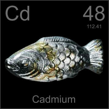
Cadmium is a known carcinogen and affects the cardiovascular, reproductive, respiratory and gastrointestinal systems, and has many detrimental effects on development. Cadmium damages the kidneys, lungs, and bones. Long-term exposure to lower levels of cadmium in air, food, or water leads to a buildup of cadmium in the kidneys and possible kidney disease. Other long-term effects are lung damage and fragile bones.
Cadmium and cadmium compounds are known carcinogens. It causes a reduction in the production of the critical white blood cells (T-Lymphocytes) which defend the body by destroying harmful free radicals and cancer cells. A few studies in animals indicate that younger animals absorb more cadmium than adults. Animal studies also indicate that the young are more susceptible than adults to a loss of bone and decreased bone strength from exposure to cadmium. In laboratory animals, cadmium causes decreases in fetal or pup body weight, skeletal malformations, and behavioral alterations. The list of diseases that cadmium can promote is extensive, including cancer, diabetes, arthritis, cardiovascular disease, kidney disease, bone disorders, and digestive problems.
Symptoms of high tissue cadmium levels include fatigue, hair loss, increased susceptibility to infection, slow healing of wounds, skin lesions, loss of smell, yellow coloration of teeth, inflammation of mucous membrane of the nose, and loss of appetite.
Cadmium is used in the production of many industrial products, including batteries, pigments, metal coatings, and plastics. We are exposed by breathing secondhand smoke or living with a smoker. Cadmium aerosols with small particle sizes, such as found in cigarette smoke, is more absorbed than larger particle sizes. Cadmium contaminated foods are another source. Low levels are found in all foods, highest levels are found in shellfish, liver and kidney meats. Cadmium has been found in at least 1,014 of the 1,669 National Priorities List sites identified by the Environmental Protection Agency.
Cadmium antagonizes and prevents the absorption of important nutrient minerals such as zinc and sulfur. Cadmium displaces zinc, which leads to zinc deficiency, resulting in slow wound healing, premature aging and reproductive problems. Since most commercial dog foods are low in zinc, this is a big concern. Zinc is a component of many vital enzymes promoting a healthy immune system, liver, and bones. Adequate zinc levels will inhibit absorption and retention of cadmium.
An hTMA analysis will verify what your tissue cadmium and zinc levels are and whether there are adequate amounts of the protective minerals present to prevent cadmium accumulation in the tissues.
Biomonitoring - Testing for Heavy Metals
Clinically, metal "toxicity" is typically designated by the measurable amount above a predetermined clinical level for some heavy metals (when indicated using tissue sampling and testing). Clinical determination is based on this tissue level elevation combined with accompanying signs and symptoms related to the specific metal in question.
In contrast, a toxic burden may fall below a clinically defined limit and can exist without accompanying signs and symptoms commonly associated with that specific metal. Under these circumstances, it should be termed a burden or increased burden and should not be referred to as a toxicity. This is not to say that the particular metal is not having a metabolic impact or that it should not be addressed. You can have a heavy metal burden, while not technically a toxicity designation, and still manifest many signs and symptoms due to an adverse or allergic reaction to that metal.
Tissues used to test for toxic levels of the heavy metals in include blood, urine, hair, fingernail, and fecal samples. Most medical offices are not equipped to perform these tests and samples must be sent to appropriate laboratories that perform such testing. For measuring effects due to acute exposures within days or up to several months, blood, urine, and fecal analysis are the most accurate. For long term and cumulative body burden effects, hair and fingernail tests are most accurate.
Blood serum testing vs. hair tissue mineral analysis.
The blood and serum do contain minerals, but they may not be completely representative of the body's mineral storage. In many cases, the serum level of minerals is maintained at the expense of tissue concentration (homeostatic mechanisms). Serum concentrations may fluctuate with emotional changes, the time of day the blood is drawn or foods eaten prior to taking a sample. For example, serum magnesium can fluctuate depending upon the blood drawing technique. The longer the tourniquet is applied, the higher the magnesium rises as a result of tissue hypoxia.
Blood analysis for minerals is a good indicator of the transport of minerals to and from the storage areas of the body (extracellular).
Excess accumulation of minerals in the body are often undetected in the serum due to their removal from the blood for deposition into the tissues. When this occurs, the mineral may fail to be excreted through the urine or intestinal tract. Thirty to forty days following an acute exposure to the toxic metal lead, for instance, elevated serum levels may be undetectable as a result of the body's removing the lead from the serum as a protective measure and depositing the metal into such tissues as the liver, bones, teeth, and hair.
Minerals may fluctuate between the serum and tissues in acute or chronic conditions. This is seen with copper and iron during infections, inflammatory disorders, and certain malignancies. Also, calcium loss from the body can become so advanced that severe osteoporosis develops without any appreciable changes noted in the blood levels of calcium.
hTMA is used worldwide and is a standard toxicology screen for biomonitoring. The U.S. EPA recommends hTMA as the preferred method of testing for chronic heavy-metal exposure. Lab results determine your toxic load, and retests are used to monitor detoxification progress.
Heavy metals will often be elevated when there has been a chronic exposure. However, the hTMA can indicate the "tip of the iceberg" so to speak, in that heavy metals can be stored or sequestered in organs and tissues throughout the body. Lead and cadmium for instance are bone seekers. Eventually they will be sequestered into the bone. As bone turnover occurs, lead and cadmium will constantly be released back into circulation and continually be incorporated into the hair shaft, hTMA will reveal the chronic nature of exposure. However, with therapy aimed at properly balancing essential minerals, removal of lead and cadmium will be hastened. This will result in even higher levels on follow-up hTMA tests as greater amounts are being released from storage areas.
Advantages of hTMA
- Hair is invaluable in the assessment of toxic metal levels. (Recommended by the EPA.)
- Hair can be sampled more quickly and easily than blood, using a safe non-invasive method.
- Unlike blood, hair is less susceptible to reactive homeostatic mechanisms that affect trace element levels.
- Long-term deviations of mineral retention or losses are more easily detected in hair than blood.
- Concentrations of most elements in the hair are significantly higher than blood and other tissues.
- Hair provides a record of past as well as present trace element levels, i.e. biological activity.
- Hair provides information of substances entering from the blood serum and external sources.
- hTMA is more cost-effective than mineral testing through other means.
Toxic Elements
The toxic elements section of a hTMA report shows the results for several toxic heavy-metals. It is preferable that all levels be as low as possible and within the lower white section. Any test result that falls within the upper dark red areas should be considered as statistically significant, but not necessarily clinically significant. Further investigation may then be warranted to determine the possibility of actual clinical significance. The toxic minerals (heavy metals) are well-known for their interference upon normal biochemical function. These toxins are commonly found in the environment and therefore are present to some degree in all biological systems. However, these metals clearly pose a concern for toxicity when tissue accumulation occurs to excess.
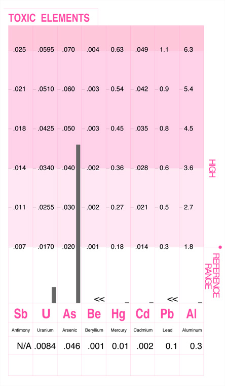
Toxic Elements: This chart shows arsenic above the established reference range. Arsenic has been found high in some seafood obtained from coastal waters, particularly shellfish. Other sources include arsenic rich soils, herbicides, arsenic containing insect sprays, burning of arsenate treated building materials in fireplaces, coal combustion, and smelters.
Toxic Ratios
This section displays the relationships between the important nutritional elements and toxic metals. Each toxic metal ratio result should be in the upper white area of the graph, and the higher the better. Toxic ratios that fall within the darker red area may indicate an interference of that toxic metal upon the utilization of the nutritional element.
Every person is exposed to toxic metals to some degree. The retention of these toxic metals, however, is dependent upon the individual's susceptibility. The balance of the protective nutrient minerals within the body in relation to the heavy metals can frequently be the determining factor to this susceptibility. As an example, the accumulation of lead will have a more detrimental effect upon body chemistry when sufficient levels of calcium and iron are not available. By examining the toxic metal levels in relation to the protective minerals, the extent to which the heavy metals may be involved in abnormal chemistry can frequently be seen. This is done by examining the toxic ratios.
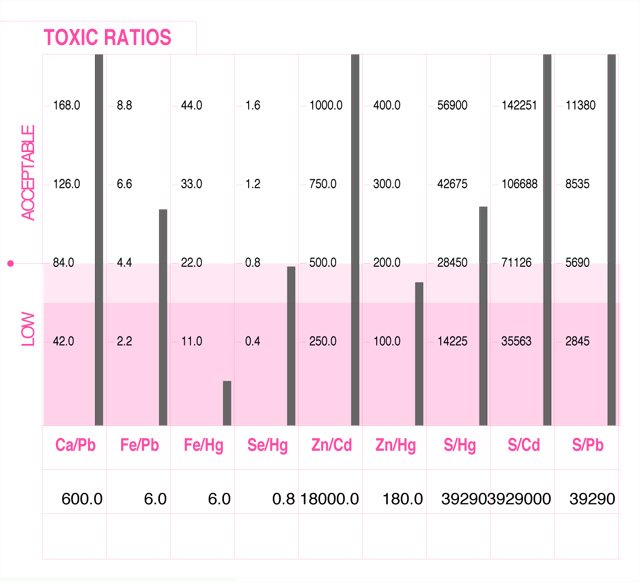
Toxic Ratios: This chart shows both a low Fe/Hg and a low Zn/Hg level. When iron and zinc levels within the body are sufficient, these nutrient minerals are able to produce an antagonistic or protective response to the adverse affects of mercury. However, when iron or zinc are low in relation to mercury, their protective action upon mercury may become markedly reduced.
Back to TopResolving Toxicity
Chelation therapy is a common prescription for detoxification from heavy metal poisoning. Chelation therapy is the introduction of compounds into the body to remove heavy metals by binding to the metal ions and holding them in suspension until they are excreted. Examples:
- Ethylenediaminetetraacetic acid (EDTA) is frequently used for severe cases of lead poisoning.
- Dimercaprol binds to both arsenic and mercury.
- Dimercaptosuccinic acid, or DMSA, binds to lead, mercury, and arsenic.
Chelating agents are typically administered in cases of acute toxicity. The removal of toxic heavy metals through chelation therapy can be extremely difficult for a dog's body and must be administered under the care of a qualified physician familiar with the process of toxin removal. Tests and therapy can be expensive and risky, progress of the therapy must be properly monitored to keep an eye out for signs of complications.
Chelation therapy can be an effective, and often necessary treatment for acute poisoning. But what about therapy for chronic exposure to toxins that you face every day? hTMA is effective for protection against persistent, low-level chronic exposure. Corrective nutritional therapy keeps the body's immune system functioning properly (allowing it to better defend itself against acute exposures as well). With hTMA, the use of harsh chelation therapy is unnecessary. Evidence now shows that even low lead levels can adversely affect neurological function. Low mercury levels can affect fetal development. All of these toxic metals have adverse effects at low levels. hTMA is an effective screening tool for persistent low-level exposures.
Since poor diet is associated with higher heavy metal accumulation, improved nutrition can relieve and prevent low-level metal toxicities and the disorders that accompany them. When nutrient minerals are available at the optimal levels and ratios, they not only help protect against the absorption of toxic metals, they also hasten their removal.
Nutrients that are known to protect against heavy metal accumulation include; calcium, magnesium, iron, copper, zinc, selenium, vitamins A, C, E, B-6, B-12, pectin, lecithin, glutathione and other antioxidants. Many of the essential amino acids found in whole, unprocessed foods help also. Specialty foods like kelp, garlic, and brewer's yeast can add detoxification benefits. Providing protective nutrients in the optimal amounts and ratios will prevent you from becoming undernourished and thus susceptible to the accumulation of toxic metals in their body tissues.
hTMA provides the information needed to detect and correct mineral imbalances and optimize metabolism. Follow-up lab tests allow you to monitor the progress of toxin removal. Resolving toxicity related pathologies is complex and may be dangerous. We recommend you work closely with a qualified hTMA practitioner.
Back to TopResources
- Is the World Making You Sick? Jill Neimark, interview with immunologist Claudia Miller.
- Are You Crooked? Man-Made Disease Explained by Forrest Maready.
-
Toxicant-Induced Loss of Tolerance (TILT) Self-Test Screening Forms
Form #1 (download below) is a 3-question screening tool to help you determine if you should complete form #2 (download below) Quick Environmental Exposure and Sensitivity Inventory. TILT research.org
- Aluminum Toxicity in Infants and Children American Academy of Pediatrics, 1996.
- Hair Analysis and Heavy Metals - Hidden Metal Detox
- A Small Dose of Toxicology Steven Gilbert
- Implications of Lead Toxicity
- Quick FAQ - Aluminum
- Quick FAQ - Arsenic
- Quick FAQ - Cadmium
- Quick FAQ - Lead
- Quick FAQ - Mercury
- The Poison Papers Compilation of over 20,000 chemical industry and regulatory agency documents and correspondence stretching back to the 1920s. Proof that both industry and regulators understood the extraordinary toxicity of many chemical products and worked together to conceal this information. They expose the hazards posed by certain chemicals and the fraudulence of some regulatory processes relied upon to protect human health and the environment.
- Toxic Cocktail: How Chemical Pollution Is Poisoning Our Brains Barbara Demeneix
- Mental and Elemental Nutrients Carl. C. Pfeiffer, MD, Ph.D.
- Trace Elements in Human and Animal Nutrition U.S. Dept. of Agriculture
- CDC - Agency for Toxic Substances and Disease Registry
- CDC: Protecting the Private Good? Marcia Angell, former editor in chief of the New England Journal of Medicine, explains, "The CDC has enormous credibility among physicians, in no small part because the agency is generally thought to be free of industry bias. Financial dealings with biopharmaceutical companies threaten that reputation."